Leg Bone Diagram - Bones Human Anatomy Organs / Its lower end helps create the knee joint.. At the same time, the bones and joints of the leg and foot must be strong enough to support the body's weight while remaining. Posted on april 18, 2019april 18, 2019. Joints of hand anterior view, lateral view, right hand. Bone diagram forehead (frontal bone) nose bones (nasals) cheek bone (zygoma) upper jaw (maxilla) lower jaw (mandible) breast bone (sternum) upper arm bone (humerus) lower arm bone (ulna) thigh bone (femur) collar bone (clavicle) toe bones (phalanges) ankle bones (tarsals) kneecap (patella) shin bone 10 / 10 ( 1 vote ) leg bone anatomy diagram diagram of human leg human anatomy diagram.
Lower jaw (mandible) collar bone. Dog leg anatomy is complex, especially dog knees, which are found on the hind legs. The hip itself is a ball and socket joint, much like the shoulder.the structures necessary to create this joint are the socket, the joint capsule, muscle, ligaments, and the neck. It is the largest bone in the body and is the only bone in the upper leg. Some types of leg pain can be traced to problems in your lower spine.
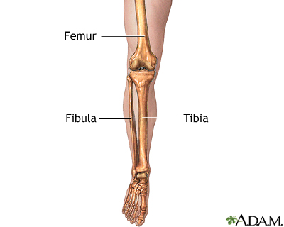
Leg Skeletal Anatomy Medlineplus Medical Encyclopedia Image from medlineplus.gov The foot bones shown in this diagram are the talus, navicular, cuneiform, cuboid, metatarsals and. The bones together make up the hip. The lower leg extends from the knee to the ankle. The technical term for a dog knee is the stifle joint. The pubis, ischium, and ilium together constitute the pelvis while the thigh bone is the femur. Ankle bones anatomy, arm bones anatomy, fibula anatomy, fibula fracture, hip bones anatomy, leg bones human body, foot, ankle bones anatomy, arm bones anatomy, fibula anatomy, fibula fracture, hip bones anatomy, leg bones human body. These muscles work together to produce movements such as standing, walking, running, and jumping. In this image, you will find femur, medial epicondyle of the femur, patella, tibial tuberosity, anterior rest of the tibia, a medial surface of the tibia, lateral epicondyle of the femur, head of the fibula, fibula, medial malleolus of the tibia, lateral.
Its lower end helps create the knee joint.
15 photos of the leg bones anatomy diagram. The stifle joint connects the femur, which is the dog thigh bone, to the tibia and fibula, the lower leg bones, and the patella,the canine equivalent to the knee cap. Lower jaw (mandible) collar bone. The rounded, proximal end is the head of the femur, which articulates with the acetabulum of the hip bone to form the hip joint. The knee joint is the largest joint in the body and is primarily a hinge joint, although some sliding and rotation occur. Also called the shin bone, the tibia is the longer of the two bones in the. The femur, or thighbone, is the longest and largest bone in the human body. The lower leg extends from the knee to the ankle. The femur, or thigh bone, is the single bone of the thigh region (figure 6.51). High resolution textures and displacement included. Bone diagram forehead (frontal bone) nose bones (nasals) cheek bone (zygoma) upper jaw (maxilla) lower jaw (mandible) breast bone (sternum) upper arm bone (humerus) lower arm bone (ulna) thigh bone (femur) collar bone (clavicle) toe bones (phalanges) ankle bones (tarsals) kneecap (patella) shin bone Long bones, short bones, flat bones, and irregular bones.) long bones are longer than they are wide, with spongy bones at both ends and a cavity filled with bone marrow in the shaft. The tibia and fibula are two long bones that run parallel to each other, forming the scaffold of the leg and providing attachment points for many muscles.
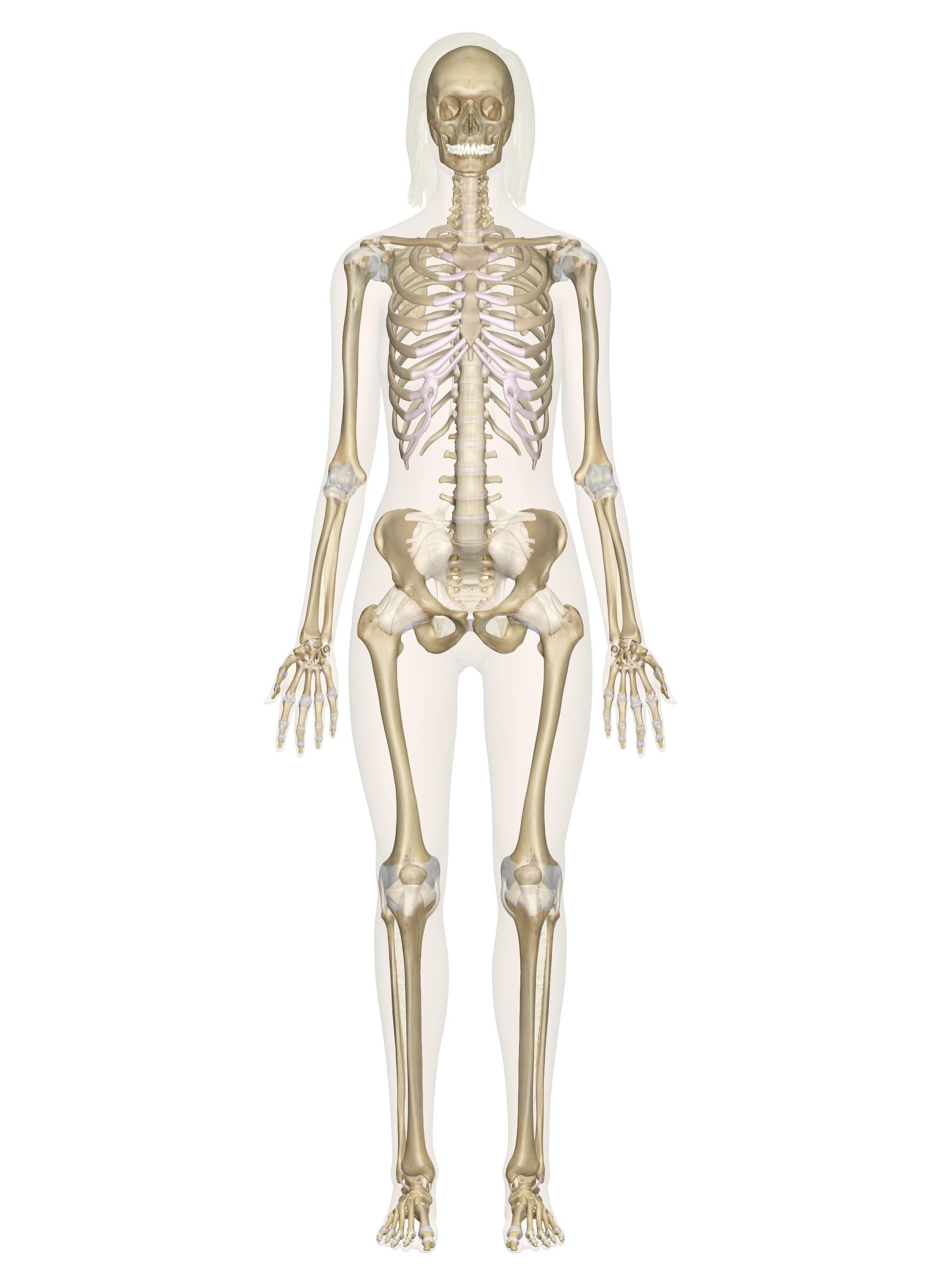
(there are four types of bone: The tibia, commonly known as the 'shin bone', is the largest and most medial of the two.you can palpate its anterior border when you run your finger down the anterior aspect of your leg. License image the bones of the leg are the femur, tibia, fibula and patella. The bones of the leg and foot form part of the appendicular skeleton that supports the many muscles of the lower limbs. These bones are arranged into two major divisions:
Practical Art Anatomy E G Lutz from drawingbooks.org License image the bones of the leg are the femur, tibia, fibula and patella. The knee joint is the largest joint in the body and is primarily a hinge joint, although some sliding and rotation occur. These bones are arranged into two major divisions: The hip bone (os coxae, innominate bone, pelvic bone or coxal bone) is a large irregular bone, constricted in the center and expanded above and below leg bone diagram. Also called the shin bone, the tibia is the longer of the two bones in the. Lower jaw (mandible) collar bone. Blood vessels and nerves enter the bone. The foot bones shown in this diagram are the talus, navicular, cuneiform, cuboid, metatarsals and calcaneus.
It is the largest bone in the body and is the only bone in the upper leg.
The bones of the leg and foot form part of the appendicular skeleton that supports the many muscles of the lower limbs. The lower leg extends from the knee to the ankle. Dog leg bone diagram / dog anatomy leg bones stock image stock photo download image now istock / paw bone between the heel and the phalanges.license image the bones of the leg are the femur, tibia, fibula and the foot bones shown in this diagram are the talus, navicular, cuneiform, cuboid, metatarsals and from dogs with three legs to cats without eyes, the perfect imperfection photo series. 15 photos of the leg bones anatomy diagram. The bones of the leg are the femur, tibia, fibula and patella.the foot bones shown in this diagram are the talus, navicular, cuneiform, cuboid, metatarsals and calcaneus. Some types of leg pain can be traced to problems in your lower spine. The femur is known as a long bone. Dog leg anatomy is complex, especially dog knees, which are found on the hind legs. These bones have a marrow, but not a bone marrow cavity. Also called the shin bone, the tibia is the longer of the two bones in the. Its lower end helps create the knee joint. The rounded, proximal end is the head of the femur, which articulates with the acetabulum of the hip bone to form the hip joint. Disposition of rotator cuff muscles diagram.
Related posts of diagram of leg bones long bone femur label. Hip and leg bone diagram / lower leg bones anatomy anatomy drawing diagram / this lengthy bone connects with the knee at one finish and the ankle on the different. The bones together make up the hip. Its lower end helps create the knee joint. The hip itself is a ball and socket joint, much like the shoulder.the structures necessary to create this joint are the socket, the joint capsule, muscle, ligaments, and the neck.
Skeletal System Labeled Diagrams Of The Human Skeleton from innerbody.imgix.net Electrical wiring diagrams leg bones diagram femur which are in coloration have a bonus above when looking at any leg bones diagram femur wiring diagram, get started by familiarizing your self. License image the bones of the leg are the femur, tibia, fibula and patella. At the same time, the bones and joints of the leg and foot must be strong enough to support the body's weight while remaining. With different grades of sprains depending on severity. Dog leg anatomy is complex, especially dog knees, which are found on the hind legs. Its lower end helps create the knee joint. The medial, larger bone of the lower leg. The pubis, ischium, and ilium together constitute the pelvis while the thigh bone is the femur.
Disposition of rotator cuff muscles diagram.
Dog leg anatomy is complex, especially dog knees, which are found on the hind legs. The bones of the leg are the femur, tibia, fibula and patella.the foot bones shown in this diagram are the talus, navicular, cuneiform, cuboid, metatarsals and calcaneus. The fibula is connected via ligaments. With different grades of sprains depending on severity. Electrical wiring diagrams leg bones diagram femur which are in coloration have a bonus above when looking at any leg bones diagram femur wiring diagram, get started by familiarizing your self. The technical term for a dog knee is the stifle joint. Also called the shin bone, the tibia is the longer of the two bones in the. Related posts of diagram of leg bones long bone femur label. Leg pain can also be caused by blood clots, varicose veins or poor circulation. The proximal portion of the tibia is tibial plateau which acts as a cusp for the knee, the distal portion tapers into the medial malleoli and the concave surface which articulates with the talus at the ankle joint. Blood vessels and nerves enter the bone. Each leg is made up of four bones. The tibia and the fibula, at the top of the ankle joint.